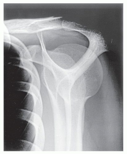
Outlet View Of Shoulder Joint X-ray


Shoulder Arthritis / Rotator Cuff Tears: causes of ...
Let's take a look at how the shoulder should be x-rayed to diagnose shoulder arthritis and to plan a joint replacement. In the shoulder, two X-ray views are essential for determining the 'joint space', the AP (anteroposterior) and the axillary views. An AP view of a normal shoulder looks like this:Supraspinatus Outlet view Shoulder Xray. Demonstrates ...
Aug 15, 2014 - Supraspinatus Outlet view Shoulder Xray. Demonstrates: outlet/impingement of the supraspinatus and coracoacromial arch.Y view
The anteroposterior view with the arm at 30 degrees external rotation, the outlet Y view and the axillary view. The outlet Y view is useful because it shows the subacromial space and can differentiate the acromion processes. A lateral outlet view of shoulder joint x-ray view of the scapula will also do this. With shoulder impingment you certainly want to examine the acromion. tas lv neverfull
Jan 27, 2012 · Scapula Lateral (Y) view 5. Routine (transthoracic) AP view of the shoulder• AP relative to thorax• Suboptimal view of glenohumeral joint• Good view of AC joint 6. Scapular/Glenohumeral AP view (aka Oblique view)• Better visualize Glenohumeral joint/space• Suboptimal view of AC joint 7.
The glenohumearal joint has a greater range of motion than any other joint in the body. The small size of the glenoid fossa and the outlet view of shoulder joint x-ray relative laxity of the joint capsule renders the joint relatively unstable and prone to subluxation and dislocation. MR is the best imaging modality to examen patients with shoulder …
Shoulder XRay - www.bagssaleusa.com
Signs of prior Anterior Shoulder Dislocation. Hill-Sachs Lesion (posterior humeral head indentation) Impact occurs when Shoulder dislocates anterior to glenoid; Signs of Osteoarthritis. Axillary view best demonstrates joint space narrowing; Subchondral sclerosis and osteophytes may also be seenTRANSTHORACIC LATERAL SHOULDER (TRAUMA) - …
Apr 15, 2012 · X-ray examination of proximal humerus (shoulder) in lateral view transthoracic. This view is also know as the Lawrence Method, to demonstrate the proximal humerus. A breathing technique is done to blur the overlying ribs and lung markings. Performing this technique and prevented of patient motion the humerus should appear sharp.Conventional Radiography of the Shoulder
Figure 2 (A) Grade I sprain of AC joint. AP view of shoulder in patient with pain and point tenderness of AC joint after fall onto shoulder demonstrates normal appearing AC joint (arrow) with no separation or fracture. (B) Grade II sprain of AC joint. AP outlet view of shoulder joint x-ray view of shoulder demonstrates slight …RECENT POSTS:
- louis vuitton popincourt mm bag
- louis vuitton sneakers homme 2019-20
- louis damier azur
- youtube louis vuitton favorite pm and mm
- lv mini backpack bracelet
- mens dress shoes on sale at macys
- louis vuitton store opening san antonio
- lv wallets small
- louis martini ice bag
- louis vuitton polo shirt long sleeved
- neverfull mm or neverfull gm
- louis vuitton vernis alma gm xl
- inside a real lv bag
- lv x off white bag
All in all, I'm obsessed with my new bag. My Neverfull GM came in looking pristine (even better than the Fashionphile description) and I use it - no joke - weekly.
Other handbag blog posts I've written:
tote bag with zipper pattern free
yest saint laurent outlet store
how to say louis vuitton in spanish
louis vuitton noe purse m57099
louis vuitton mini pochette sold outside
Do you have the Neverfull GM? Do you shop pre-loved? Share your tips and tricks in the comments below!
*Blondes & Bagels uses affiliate links. Please read the louis vuitton travel guide paris for more info.