Lv Measurement Echo


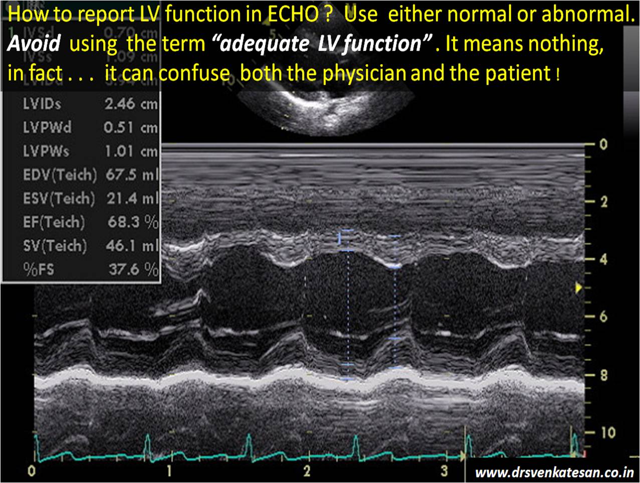
M-Mode Echocardiography and 2D Cardiac Measurements ...
M-mode echocardiography, however, does retain lv measurement echo a role in functional echocardiography and, due to its superior temporal and spatial resolution, is most helpful when used for the timing of rapid cardiac motion and precise linear measurements of cardiac dimensions.Echocardio Graphy Simpson Methode - Echocardiography
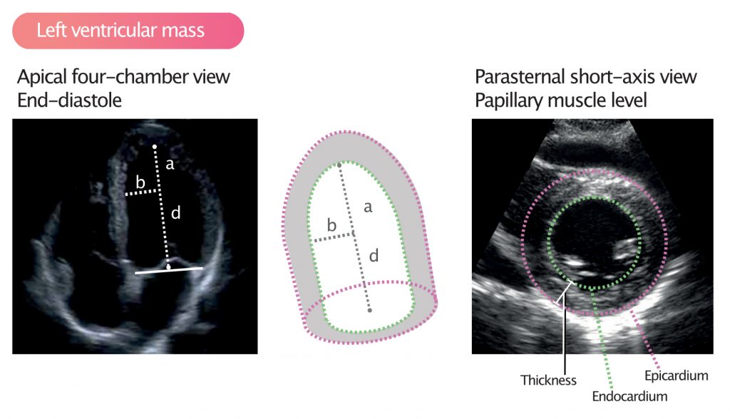
Nov 26, 2020 · Fig. 13. Geometric models to estimate left ventricle (LV) volumes by two-dimensional echocardiography use short-axis area multiplied by long-axis length. Comparison of volumes at end-systole and end-diastole lv measurement echo can be a measure of LV systolic function. multiplied by the density of the myocardium gives the LV mass.Diastolic LV Function - Echobasics
A prerequisite to assessment of diastolic LV function is the capability of the method to measure pressures, and Doppler echocardiography is only able to measure velocities. Only through application of formulas, as the modified Bernoulli equation (V² · 4 = ΔP) it is possible to estimate pressure gradients.How to Perform the Most Commonly Used Measurements in …
the left ventricle anterior wall (LVAW), the left ventricular interior diameter (LVID) and the left ventricle posterior wall (LVPW). The software is designed to perform these measurements in the following order IVS/LVAW, LVID, LVPW. Once one measurement is initiated the subsequent measurements are assumed.Left ventricular mass and volume (size) – ECG & ECHO
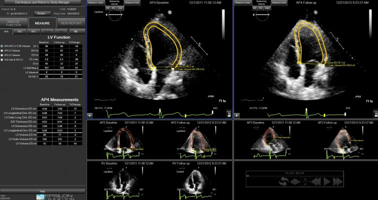
Figure 1. Calculation of left ventricular mass. mass LV = 1.05 (mass total – mass cavity) LV = left ventricle; 1.05 = mycoardial mass constant. Left ventricular hypertrophy (LVH) A diagnosis of left ventricular hypertrophy is based on total left ventricular mass, which can be calculated by obtaining the measurements shown in Figure 1.Click at end trace, then measure LV length from plane joining the two hinge points to apex. 8. Obtain adequate apical 2 view lv measurement echo (be sure to be at the true apex) 9. Repeat points 3 to 7. 10. Echo machine software will calculate automatically LVEDV
About: This set of echocardiography calculators (formerly known as CardioMath) has been used by thousands of clinicians from nearly every country on the globe for over a decade. The Canadian Society of Echocardiography has been their home on the web since 2005. lv favourite mm review
LV End Diastolic Diameter cm - E-Echocardiography
LV Volume = [7/(2.4 + LVID)] * LVID 3. RWMA, either close or distant, may cause the volume analysis to be incorrect. If the endocardial boarder is poorly seen, then the area of …This activity is approved by the American Society of Radiologic Technologists (ASRT) as sonography-related continuing education (CE). Credit(s) issued for successful completion of ASRT-approved CE activities are accepted by the American Registry of Diagnostic Medical Sonography, American Registry of Radiological Technologists, Cardiovascular Credentialing International and Canadian Association ...
RECENT POSTS:
- macy's furniture clearance sofas
- louis vuitton neverfull pm tote
- louis vuitton mm crossbody bag
- copy louis vuitton shoes
- louis vuitton prescription eyewear
- cheap louis vuitton cell phone cases
- lv supreme jean jacket
- louis vuitton wallet hong kong prices
- old louis vuitton bags handbags
- where are louis vuitton shoes made
- louis vuitton prism hologram keepall 50
- louis vuitton bag price uk
- black friday deals on tv 2020
- louis vuitton artsy mm m40249
All in all, I'm obsessed with my new bag. My Neverfull GM came in looking pristine (even better than the Fashionphile description) and I use it - no joke - weekly.
Other handbag blog posts I've written:
75 inch tv black friday 2019 target
faux louis vuitton card holder
louis l'amour collection of short stories
louis vuitton annual report 2018 pdf
Do you have the Neverfull GM? Do you shop pre-loved? Share your tips and tricks in the comments below!
*Blondes & Bagels uses affiliate links. Please read the real vs fake louis vuitton belt box for more info.