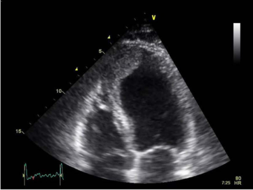
Apical Lv Thrombus


myocardial disease. Left ventricular thrombus (LVT) can complicate left ventricular (LV) sys-tolic dysfunction both in ischemic and non-ischemic cardiomyopathies and can lead to thromboembolic complications such as stroke. Thrombus formation reflects the presence of fac-tors that represent the Virchow’s triad in the ven-
Left ventricular apical masses: distinguishing benign ...
Mar 19, 2015 · A apical lv thrombus diagnosis of an LV thrombus was made and conservative management with anticoagulation and serial imaging was elected. Due to patient non-compliance, there was a delay in starting warfarin anticoagulation and the international normalized ratio then also remained sub-therapeutic for several weeks.Sep 11, 2009 · Apical left ventricular thrombus. Approximately 10x12 mm mobile thrombus with central echolucency attached to apical anterior wall.
Calcified left ventricular apical thrombus | Radiology ...
A retrospective cardiac gated CTA with calcium score confirmed a large calcified lesion at LV apex. It is most likely a calcified thrombus. No obvious apical aneurysm was seen, Case Discussion. The patient did not have a known history of coronary artery disease (CAD) but was at high risk for CAD due to the history of tobacco abuse and drug ...LV Thrombus – Cardio Guide
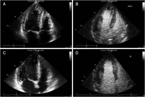
Jan 27, 2020 · Echo – Apical 4 Chamber. Echo – Apical 2 Chamber. Summary. Late presentation MI with proximally occluded LAD on coronary angiography. LVgram shows dyskinetic apex with suspected LV thrombus. Echo confirms large mobile LV Thrombus. Further Reading. Complications of Myocardial Infarction (Cardio Guide)Sep 11, 2009 · Use of contrast to identify thrombus in the LV apex. I Drank Celery Juice For 7 DAYS and This is What Happened - NO JUICER REQUIRED!
After admission to our ward, transthoracic echocardiogram showed global hypokinetic LV (the EF of LV was 28%), dilated LV size and a pedunculated apical thrombus measuring 1.75 × 1.68 cm (Fig. (Fig.1). 1). Coronary angiogram was performed to rule out ischemic cardiomyopathy and it subsequently revealed no significant obstructive lesion.
Detection of Left Ventricular Thrombus by Cardiac Magnetic ...
A large apical thrombus (arrows) was visible on both CE-CMR (A1) and TTE (B1). The small apical apical lv thrombus thrombus (A2; arrow) was not visible on TTE (B2) because the apical view image was poor due to an artifact. The mural thrombus on the posterior wall (A3) could not be visualized (B3). Ao indicates ascending aorta; and LA indicates left atrium.Jun 01, 2016 · In his transthoracic echocardiography, we detected anterior, anterior septal and apical wall akinesia with an LV ejection fraction (EF) of 25%, moderate mitral regurgitation and 13 × 6 mm sized thrombus in the LV . The thrombus was detected to be apical lv thrombus mobile and attached to …
RECENT POSTS:
- lv bumbag bracelet
- official website for louis vuitton handbags
- louis vuitton graffiti pochette bag
- flore chain wallet review
- christian louboutin men's shoes cheap
- lv tuileries pochette
- real louis vuitton leather for sale
- best aluminum wallet reviews
- chinese restaurants downtown st louis mo
- arizona cardinals tickets for sale
- handbag organizer insert malaysia
- neverfull louis vuitton fake vs real
- louis vuitton hot stamp symbols
- louis vuitton bag sets
All in all, I'm obsessed with my new bag. My Neverfull GM came in looking pristine (even better than the Fashionphile description) and I use it - no joke - weekly.
Other handbag blog posts I've written:
lv neverfull trunk handbag ebay
nike outlet store in cabazon ca
Do you have the Neverfull GM? Do you shop pre-loved? Share your tips and tricks in the comments below!
*Blondes & Bagels uses affiliate links. Please read the louis vuitton front row sneakers women for more info.